![]()
![]()
A dual-modality intravascular imaging catheter that helps you identify lipid-core plaques that is at an increased risk of major adverse cardiac events.
Test your image interpretation skills by downloading the free IVUSAID app.
Dualpro® IVUS+NIRS is a dual-modality intravascular imaging catheter equipped with Near-Infrared Spectroscopy (NIRS) delivering intel on plaque composition and giving you the power to make highly informed decisions right in the Cath lab. Now you can easily identify unstable lipid core plaque (LCP) – a well-documented culprit in heart disease associated with 95% of STEMIs and an increased risk of peri-procedural complications.3,4
With the power of Dualpro® IVUS+NIRS, you can identify patients and plaque at a higher risk of MACE and readily distinguish between stable plaque and vulnerable LCP.
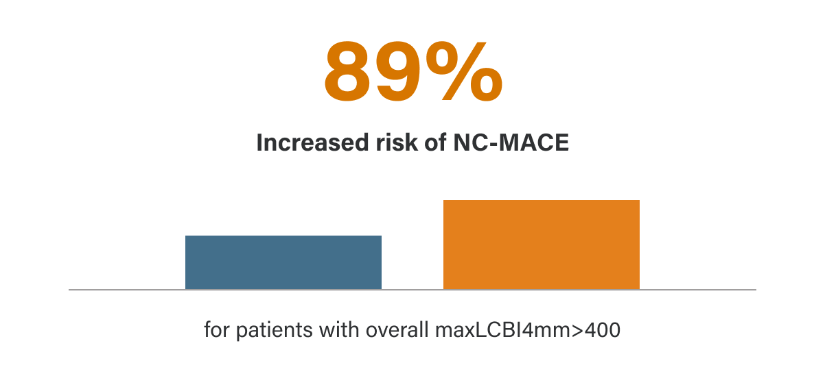
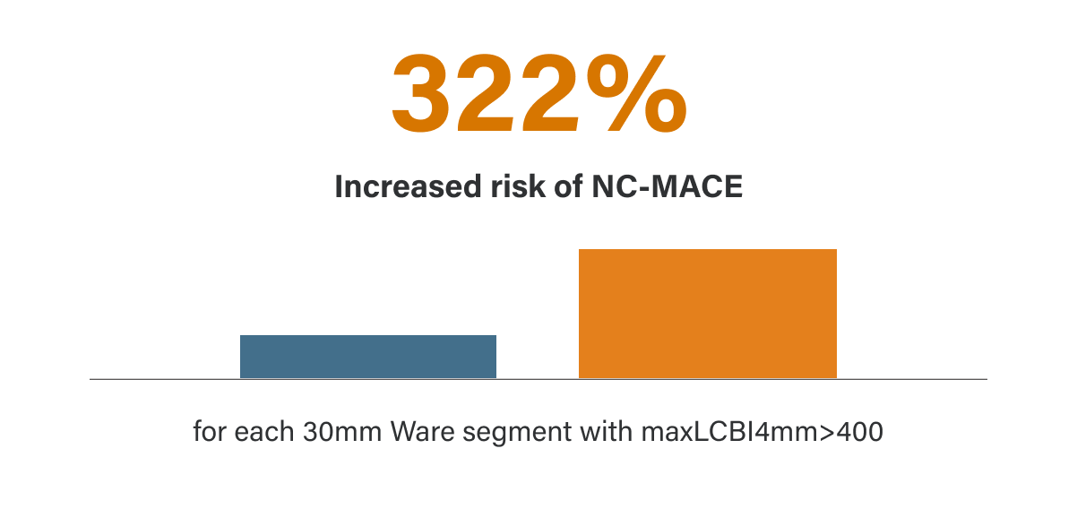
Multiple studies have demonstrated that assessment of the coronary artery with the Makoto® IVUS+NIRS Intravascular Imaging System is unmatched in its ability to identify the risk of MACE. The results of the landmark LP study provided clinical evidence that intravascular NIRS imaging can accurately identify both vulnerable patients and vulnerable plaques that are at significantly higher risk for subsequent NC-MACE during a 24-month period.5
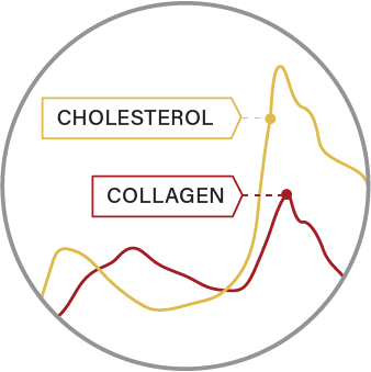
To identify lipids such as cholesterol
Organic molecules have unique spectroscopic signatures that can be used to detect their presence in a mixture of unknown composition. NIRS allows us to distinguish molecules, such as collagen and cholesterol, within the vessel wall and thus identify the presence of LCP.
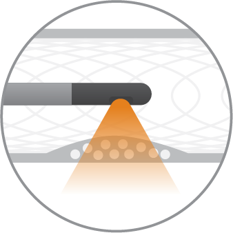
Through blood, tissue and interstitial spaces
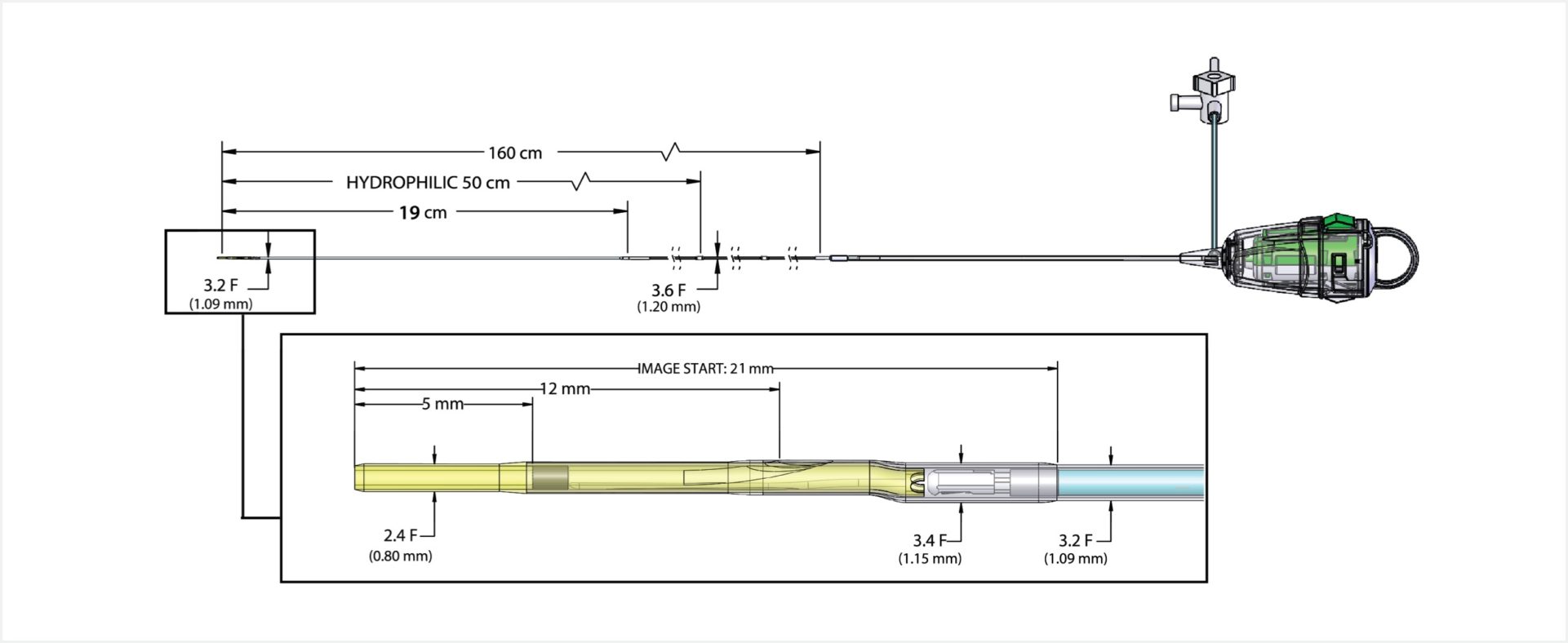
The microscopic mirrors at the tip of the Dualpro® catheter are designed to deliver near-infrared light to the vessel wall and collect the diffusely reflected light. The light propagates through blood and tissue by scattering and absorption, even in the presence of calcium or stents, to interrogate the plaque for its chemical fingerprint.
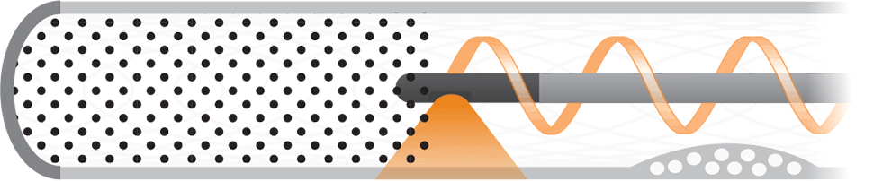
With the aid of advanced algorithms
Advanced algorithms analyze the returned light and calculate the probability of the presence of a lipid core plaque. Our algorithms have been validated in a large prospective histology study providing you with information you can trust.
The Makoto® Intravascular Imaging System was designed with the primary goal of helping you make quick and informed decisions at the bedside. The easy-to-interpret chemogram provides insight into the complex plaques that complicate your interventional strategy, treatment procedures, and patient’s recovery.
Approximately 1,300 NIRS spectra per millimeter are acquired as the catheter scans the vessel.¹
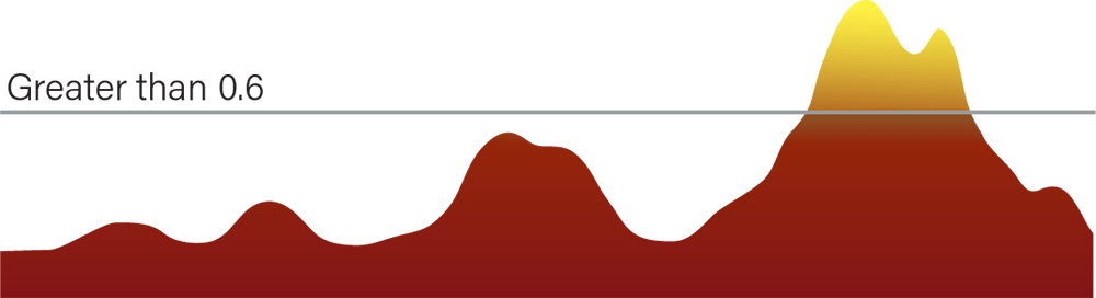
The acquired NIRS signals are analyzed and each spectrum is assigned a probability score, from 0 to 1, based on the likelihood of the presence of LCP.

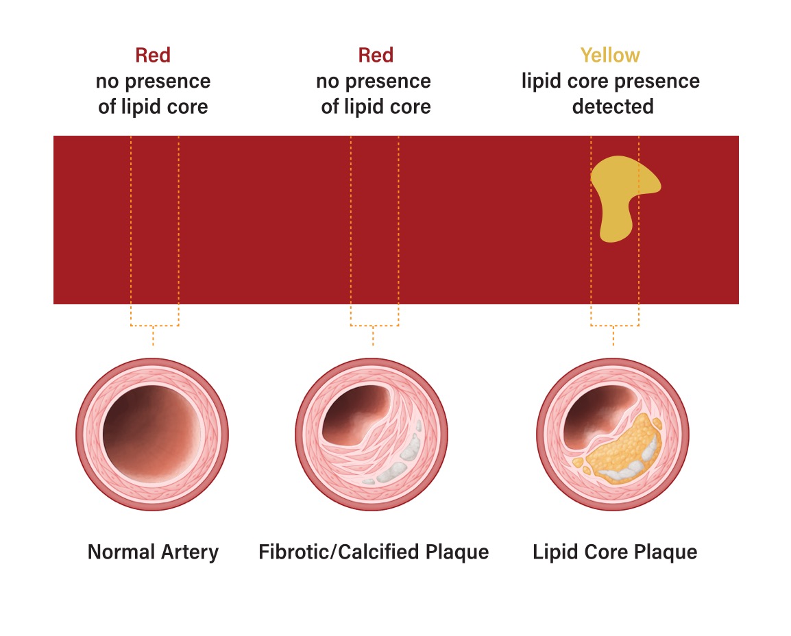
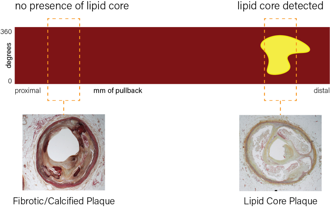
All probability scores, low to high, are mapped on a continuous color scale from red to yellow. Scores above 0.6 appear orange to yellow in the chemogram and contribute to the Lipid Core Burden Index (LCBI).
The chemogram is automatically generated within seconds, creating a map of the LCP location within the vessel wall. This color-coded map can be interpreted quickly, permitting informed treatment decisions.
Data you can trust:
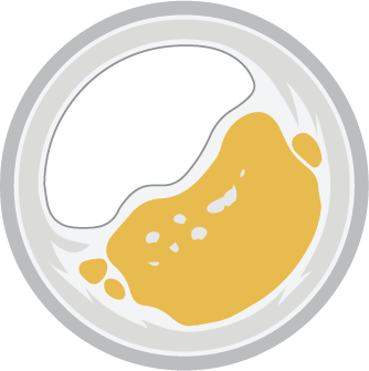
Nearly 2,500 artery cross-sections were histologically and spectrally analyzed to validate lipid core plaque detection by NIRS. The red and yellow colors on the chemogram help differentiate normal or fibrotic plaque that is presumed to be stable (left) from those that contain lipid core plaques (right).²
¹ In a 150mm scan at 05.mm/s
² Detection of lipid core coronary plaques in autopsy specimens with a novel catheter-based near-infrared spectroscopy system, Gardner er al, JACC Cardiovasc Imaging, 2008

*The Makoto Intravascular Imaging System® is intended for ultrasound examination of coronary and peripheral intravascular pathology. Intravascular ultrasound imaging is indicated in patients who are candidates for transluminal coronary and peripheral interventional procedures. The System is not indicated for use in the cerebral vessels. See product instructions for full product description, contraindications, warnings, and instructions.
US Customers: Click here to proceed to Nipro.com
References
© 2026 Infraredx™, Inc. All Rights Reserved Powered by Bloom Creative